New Patients
Existing Patients
New Patients
Existing Patients
New Patients
Existing Patients
New Patients
Existing Patients
New Patients
Existing Patients
New Patients
Existing Patients
New Patients
Existing Patients
New Patients
Existing Patients

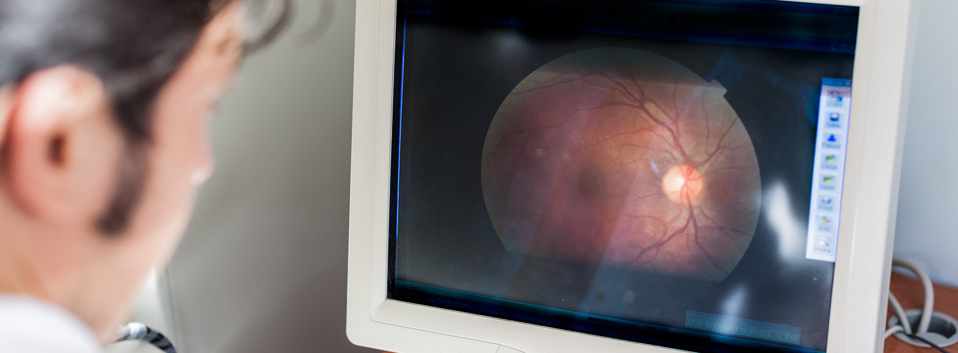
The Retinal Fundus Exam, often referred to as a Dilated Fundus Exam (DFE), is a critical component of a comprehensive eye examination. It is a procedure where an eye care professional (such as an optometrist or ophthalmologist) thoroughly inspects the back interior of your eye, known as the fundus.
The fundus is the inner back surface of the eye, containing three essential structures:
The Retina: The light-sensitive tissue that lines the back of the eye. It converts light into electrical signals that are sent to the brain.
The Optic Nerve Head (Optic Disc): The beginning of the optic nerve, which transmits visual information from the retina to the brain.
Blood Vessels: The arteries and veins that supply oxygen and nutrients to the retina.
Because the pupil (the black center of the eye) is small, it normally limits the doctor's view. To get a clear, panoramic, and three-dimensional view of these vital structures, special eye drops are used to temporarily dilate (widen) the pupil.
The DFE is considered the "gold standard" for diagnosing many serious eye and systemic diseases because it allows the doctor to see a wider area and assess depth (stereoscopic view), which is essential for:
Comprehensive Retinal Coverage: Seeing the far edges and periphery of the retina where problems like tears or detachments often begin.
Depth Assessment: Evaluating the contour and shape of the optic nerve to check for early signs of glaucoma.
Detailed Inspection: Locating subtle damage to the blood vessels and tissue.
Dilation Drops: The doctor will place medicated eye drops into your eyes. You may feel a slight stinging sensation for a few seconds.
Waiting Period: It typically takes 15 to 30 minutes for your pupils to fully dilate. Your vision will become blurry, especially for reading, and your eyes will become highly sensitive to light.
The Examination: Once the pupils are wide, the doctor will use a magnifying lens and a bright light (often with an instrument called an ophthalmoscope or slit lamp) to look deep into your eye. The entire exam is fast, simple, and painless.
Post-Exam Care: The effects of the dilation drops usually last 4 to 6 hours.
Do NOT Drive: You must arrange for transportation home, as your vision will be impaired.
Wear Sunglasses: Your eyes will be highly light-sensitive; wear sunglasses to protect them.
Since the eye is the only place where a doctor can non-invasively view living blood vessels and nerve tissue, the DFE can detect early signs of diseases affecting not just the eye, but the entire body.
| Ocular (Eye) Conditions | Systemic (Body) Conditions |
| Diabetic Retinopathy (damage to retinal vessels) | Diabetes (often detected before a formal diagnosis) |
| Glaucoma (damage to the optic nerve) | High Blood Pressure (hypertension) |
| Age-Related Macular Degeneration (AMD) | High Cholesterol (deposits in vessels) |
| Retinal Tears or Detachment | Stroke Risk (plaque deposits in retinal arteries) |
| Ocular Tumors/Cancers | Brain Tumors (via changes in the optic nerve) |
| Cataracts (clouding of the lens) | Multiple Sclerosis (via optic nerve inflammation) |
While recommended periodically for all patients as part of a comprehensive eye exam, a DFE is especially critical for individuals who:
Have Diabetes
Have a history of Glaucoma or Macular Degeneration
Are Over the age of 60
Have High Myopia (severe nearsightedness)
Have a family history of retinal or optic nerve disease

Quick Links
Contact Us