New Patients
Existing Patients
New Patients
Existing Patients
New Patients
Existing Patients
New Patients
Existing Patients
New Patients
Existing Patients
New Patients
Existing Patients
New Patients
Existing Patients
New Patients
Existing Patients

Glaucoma is not a single disorder but a group of conditions that gradually damage the optic nerve, the vital pathway that carries visual signals from the eye to the brain. In many cases the damage begins in the peripheral visual field, producing subtle blind spots that are easy to miss until they expand. Because change is often slow and painless, many people remain unaware of early loss until it becomes more advanced.
Although elevated intraocular pressure (IOP) is a major risk factor for many forms of glaucoma, damage can occur even when pressure readings fall within a so-called normal range. That complexity is why diagnosing and managing glaucoma requires more than a single test; it calls for a careful interpretation of pressure, nerve appearance, and functional testing over time. Early recognition and consistent follow-up are the best defenses against irreversible vision loss.
Risk increases with age, a family history of glaucoma, certain ethnic backgrounds, and medical conditions such as diabetes. Some people also have anatomical differences—like a crowded drainage angle in the front of the eye—that predispose them to particular types of glaucoma. Understanding individual risk helps shape a tailored monitoring and treatment plan aimed at preserving vision for years to come.
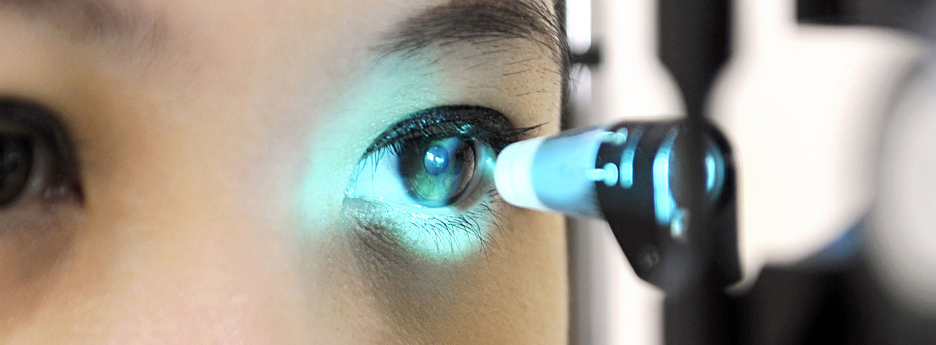
Detecting glaucoma early begins with a detailed eye exam that looks beyond a single snapshot. Our clinicians assess eye pressure but also examine the optic nerve directly, often using magnification and specialized illumination to look for subtle changes in nerve fiber loss. Evaluation of the drainage structures and the front of the eye helps identify situations that could lead to rapid pressure rises or angle-related glaucoma.
We also test visual function with perimetry, commonly called a visual field test, which maps the areas where sight may already be compromised. Repeating these tests over time lets us detect trends: a steady reduction in sensitivity in a particular area of the field is meaningful, even if absolute numbers look acceptable on one visit. Functional testing complements structural imaging to give a more complete picture.
Because glaucoma can be quiet, routine exams are essential—especially if you have risk factors. Our approach is conservative: we consider symptoms, objective findings, and the pace of any documented change before recommending a treatment plan. This careful assessment helps avoid unnecessary therapy while ensuring that those who need intervention receive it in a timely way.
Modern glaucoma management relies heavily on technology that measures both structure and function. Optical coherence tomography (OCT) provides high-resolution images of the optic nerve and the retinal nerve fiber layer, enabling detection of thinning that may precede clear field loss. Correlating OCT results with visual field testing helps clinicians separate real progression from normal variability.
Tonometry with modern devices gives an estimate of intraocular pressure, but we also consider corneal thickness and other factors that can influence readings. Gonioscopy—an examination of the drainage angle—clarifies whether the angle is open or narrow, which changes the risk profile and possible treatments. When appropriate, we may perform pachymetry, pachymetry-corrected pressure assessments, or other adjunctive testing to refine our understanding.
Regular, scheduled monitoring is a cornerstone of safe care. Because glaucoma is a chronic condition, data collected over months and years—not just a single visit—determines whether a treatment is working or needs adjustment. Our team emphasizes consistent testing intervals and clear documentation so changes are identified early and acted on promptly.
While there is no cure for glaucoma at present, numerous effective strategies exist to slow or prevent further nerve damage. First-line treatment often involves topical medications that reduce intraocular pressure either by decreasing fluid production or improving outflow. The choice of agent depends on the individual’s medical history, tolerability, and the target pressure needed to prevent progression.
In-office laser treatments are another option for many patients. Laser trabeculoplasty, for instance, can improve drainage in eyes with open-angle glaucoma and may reduce or delay the need for daily drops. For narrow-angle or angle-closure situations, laser iridotomy can relieve sudden blockages and protect the eye from acute pressure spikes. Each laser approach has a specific purpose and is selected based on the eye’s anatomy and disease behavior.
Surgical techniques are reserved for cases that do not respond to medical or laser therapy or when rapid lowering of pressure is required. Contemporary glaucoma surgeries range from minimally invasive procedures that enhance drainage with a lower risk profile to more traditional filtering surgeries that provide substantial pressure reduction. The decision to proceed with surgery follows a careful discussion of expected benefits, risks, and the patient’s daily needs and goals.
Successful glaucoma care is a partnership between the patient and the eye care team. Long-term strategies include adherence to prescribed therapies, attendance at scheduled monitoring visits, and clear communication about any new symptoms—such as sudden changes in vision, eye pain, or flashes of light. Regular assessment allows clinicians to adjust treatment targets and modalities to match how the disease is behaving.
Lifestyle measures and overall health management also play supporting roles. Controlling systemic conditions like diabetes and hypertension, avoiding unnecessary steroid use, and protecting eyes from injury are practical steps that can help preserve vision. We counsel patients on what to watch for and how to integrate treatment into their daily routines so adherence is more manageable.
At Harvard Square Eye Care we emphasize individualized care plans that balance efficacy with safety and quality of life. By combining careful monitoring, evidence-based therapies, and ongoing patient education, our goal is to maintain visual function and independence for as long as possible.
Glaucoma is a lifelong condition that requires early detection, thoughtful monitoring, and individualized treatment to minimize the risk of permanent vision loss. Advances in imaging, improved medications, and evolving surgical options give clinicians a broad toolkit for protecting sight, but success depends on regular follow-up and close communication between patient and provider.
If you have risk factors for glaucoma or are concerned about changes in your vision, please contact us to learn more about our approach and schedule an evaluation. Our team is available to discuss testing, monitoring plans, and the treatment options that may be right for you.
Glaucoma is a group of eye conditions that damage the optic nerve, the pathway that carries visual information from the eye to the brain. Damage often begins in the peripheral visual field and progresses slowly, so early loss can be easy to miss without testing. Because the condition is typically painless, routine exams are important to identify changes before they affect central vision.
Elevated intraocular pressure is a major risk factor for many types of glaucoma, but optic nerve damage can occur even when pressure readings are in a normal range. That variability makes diagnosis dependent on a combination of pressure measurements, optic nerve appearance and functional testing over time. Early recognition and consistent monitoring are the best defenses against permanent vision loss.
Risk increases with age and a family history of glaucoma, and certain ethnic backgrounds carry greater risk for specific types of the disease. Systemic conditions such as diabetes and hypertension, a history of eye injury, high myopia and long‑term steroid use can also raise the likelihood of developing glaucoma. Identifying these risk factors helps clinicians choose an appropriate schedule for testing and follow up.
Anatomical features such as a narrow drainage angle put some people at higher risk of angle‑closure glaucoma, while others are more likely to develop open‑angle disease. Knowing your personal and family eye history allows the care team to tailor monitoring and preventive recommendations. Regular comprehensive exams are especially important for people with any of these risk characteristics.
A focused glaucoma evaluation includes multiple tests that assess structure and function, not just a single measurement. Tonometry measures intraocular pressure, gonioscopy evaluates the drainage angle, and a careful optic nerve exam with magnification looks for signs of nerve fiber loss. These clinical findings are combined with objective tests to form a baseline and track change over time.
Optical coherence tomography (OCT) produces high‑resolution images of the optic nerve and retinal nerve fiber layer, while automated perimetry maps functional vision loss in the visual field. Pachymetry measures corneal thickness, which can influence how pressure readings are interpreted, and repeat testing helps distinguish true progression from normal variability. Consistent, documented testing intervals are essential for safe long‑term care.
Monitoring frequency depends on the type and severity of glaucoma, the rate of any documented change, and individual risk factors. Patients newly diagnosed or showing signs of progression may need visits every few months, while stable low‑risk patients are often seen at longer intervals such as every six to twelve months. Your clinician will recommend a personalized schedule based on test results and how the disease is behaving.
Regular follow up is critical because glaucoma is chronic and changes can be gradual but significant over time. Maintaining consistent testing at recommended intervals improves the chances of detecting progression early and adjusting treatment before vision is irreversibly affected. Clear documentation and communication between patient and provider make these comparisons meaningful.
Treatment aims to slow or halt optic nerve damage by lowering intraocular pressure, and the choice of approach is individualized. First‑line therapy commonly involves topical medications that either reduce fluid production or improve outflow; selection depends on effectiveness, tolerability and the target pressure for a given patient. Medication regimens are tailored to the patient’s overall health, ocular surface tolerance and lifestyle considerations.
Laser procedures such as selective laser trabeculoplasty can enhance drainage in open‑angle glaucoma and may reduce dependence on daily drops, while laser iridotomy prevents angle closure in narrow‑angle anatomy. When medical and laser options are insufficient, surgical procedures ranging from minimally invasive glaucoma surgeries (MIGS) to more traditional filtering operations can provide greater pressure reduction. Surgical decisions follow careful discussion of expected benefits, risks and the patient’s goals.
Your initial evaluation will include a comprehensive history and a series of tests to establish a baseline for future comparison. The visit typically involves measurement of intraocular pressure, examination of the optic nerve and retina often with dilation, gonioscopy to assess the drainage angle, and imaging such as OCT plus a visual field test. These data points provide a snapshot of structure and function that clinicians use to determine diagnosis and initial management.
After testing, the clinician will explain findings in plain language, outline recommended monitoring intervals and discuss potential treatment options if needed. Baseline documentation is crucial because future changes are identified by comparing new results to these early measurements. You should expect time for questions and a clear plan for follow up or therapy when appropriate.
Topical glaucoma medications can cause local side effects such as eye irritation, redness, or changes to the ocular surface, and some patients experience systemic effects depending on the drug class. Discussing side effects with your clinician allows consideration of alternative agents, preservative‑free formulations or changes in dosing that may improve tolerability. Never stop or change medications without consulting your eye care provider because abrupt discontinuation can increase the risk of progression.
Adherence is a major factor in successful glaucoma management, and practical strategies can help patients take drops as prescribed. Techniques include establishing a consistent routine, using reminders or dosing aids, and arranging medication reviews at visits to simplify regimens where possible. Open communication with the care team helps address barriers and ensures the treatment plan remains both effective and sustainable.
Glaucoma damage to the optic nerve is not reversible, so the focus of care is on early detection and interventions that slow or prevent further loss. Regular comprehensive eye exams are the most effective preventive measure because they can identify glaucoma in its earliest, most treatable stages. For people with known risk factors, timely monitoring and prompt treatment reduce the likelihood of significant vision impairment.
Prevention also includes managing systemic health factors such as diabetes and hypertension, avoiding unnecessary or prolonged steroid use, and protecting the eyes from injury. The practice emphasizes individualized plans that combine monitoring, patient education and risk reduction to preserve visual function for as long as possible.
Certain symptoms require prompt evaluation because they can signal an acute, sight‑threatening problem such as angle closure. Seek immediate care for sudden, severe eye pain, nausea or vomiting accompanied by abrupt vision changes, halos around lights, or a markedly red eye, as these can indicate an acute rise in eye pressure. Early treatment in these situations is essential to protect vision and relieve pain.
Other warning signs include sudden loss of vision in one eye, new flashes of light or a rapid increase in floaters, and any unexpected dramatic change in vision. If you experience these symptoms, contact your eye care provider or emergency services without delay so that appropriate testing and treatment can begin promptly.
Effective long‑term care balances the goal of preserving vision with maintaining overall quality of life through individualized targets and treatment choices. Regular monitoring, clear communication about symptoms and functional changes, and timely adjustments to therapy help people remain independent and safe in daily activities. Low‑vision resources, rehabilitation services and counseling about driving or work tasks are available when needed to support functional needs.
Lifestyle measures such as controlling systemic medical conditions, using protective eyewear to prevent injury, and avoiding inappropriate steroid use complement medical care. Consistent follow up and a collaborative relationship with the eye care team allow patients to stay informed and engaged in decisions about their care. Harvard Square Eye Care's clinicians work with each patient to create a sustainable plan that protects vision while respecting individual goals.

Quick Links
Contact Us