New Patients
Existing Patients
New Patients
Existing Patients
New Patients
Existing Patients
New Patients
Existing Patients
New Patients
Existing Patients
New Patients
Existing Patients
New Patients
Existing Patients
New Patients
Existing Patients

Clear, comfortable vision is more than a convenience — it’s a foundation for daily life. Regular, thorough eye examinations are the single best way to preserve sight, detect problems early, and keep your eyes functioning at their best. A comprehensive eye exam evaluates how well you see, how your eyes work together, and the health of the tissues inside the eye.
At Harvard Square Eye Care, we approach vision care with a blend of clinical rigor and practical attention to each patient’s needs. A full examination looks beyond the need for glasses or contact lenses to screen for conditions that may have little or no symptoms in their earliest stages. That early detection lets us manage or treat problems long before they affect quality of life.
Children, working adults, and older patients all benefit from regular exams, but the focus for each visit may differ. Whether you’re scheduling a first appointment for a preschooler, updating a contact lens fitting, or getting a baseline screening as you approach middle age, a comprehensive exam delivers targeted information about vision and ocular health.
This page explains what a comprehensive eye exam covers, how testing works, common vision problems that are detected, and why routine care matters at every stage of life. If you have questions after reading, please contact us for more information.
A comprehensive eye exam is more than a quick vision check — it is an in-depth assessment of visual function and ocular health. Beyond determining whether you need a prescription, the exam evaluates eye alignment, focusing ability, peripheral vision, and the internal structures of the eye. Many serious eye diseases begin with few obvious symptoms, so a full exam is the best way to catch them early.
During the visit we gather relevant medical history, review any symptoms, and discuss lifestyle or occupational factors that affect vision. This contextual information helps the clinician tailor the exam and interpret test results in a way that is meaningful for your life — for example, whether you spend significant time on screens, drive at night, or require precise depth perception for work or sport.
Because the eyes offer a unique view of blood vessels and nerve tissue, a careful ocular exam can also reveal signs of systemic conditions such as diabetes, high blood pressure, and certain neurological disorders. Detecting those signs allows us to coordinate care with your primary care or specialty providers when appropriate.
Finally, a comprehensive exam creates a baseline record. High-resolution images and measurements taken today can be compared to future exams to spot subtle changes over time — and that continuity of information is a key advantage in preserving vision.

A typical comprehensive eye exam is comfortable, non-invasive, and tailored to the individual. Most visits last up to an hour, though the length varies depending on age, symptoms, and whether advanced testing is needed. Early in the appointment we’ll review your medical and ocular history and discuss any visual complaints so the clinician can focus the exam on issues that matter most to you.
The clinical portion of the exam includes a series of standard, painless tests designed to measure how the eyes perform together and separately. These tests provide objective data about clarity of sight, eye movement and coordination, color perception, and the function of the pupil and optic nerve. For many patients, this set of tests is sufficient to establish a diagnosis and plan of care.
When indicated, we extend the exam with specialized imaging and measurements that give a more detailed, quantitative view of the eye’s structures. Those technologies can include retinal photography, optical coherence tomography (OCT), corneal mapping, and automated visual field testing. Images can be saved to the chart so we can track changes precisely at future visits.
Below are common tests performed during a comprehensive exam; your clinician will explain which are relevant to your visit and why.
Visual acuity testing – Measures clarity of vision for each eye and establishes whether corrective lenses are needed to reach optimal acuity.
Color vision screening – Detects hereditary color deficiencies and identifies color perception problems that can be related to retinal or optic nerve disease.
Stereopsis testing – Assesses depth perception and how well both eyes work together to form a single three-dimensional image.
Ocular motility tests – Evaluates the eye muscles and their coordination as your eyes follow or track objects in motion.
Pupil reaction testing – Observes how pupils respond to light, which provides clues about neurological function and retinal health.
Autorefraction – A computerized screening that offers an initial estimate of your prescription before the clinician refines it manually.
Retinoscopy – Uses reflected light to help estimate the corrective power needed, especially useful for pediatric patients or those unable to provide subjective feedback.
Refraction – The subjective portion of the exam where the clinician determines the exact eyeglass prescription through careful refinement and patient responses.
Keratometry – Measures the curvature of the cornea, information that is essential for contact lens fittings and some surgical planning.
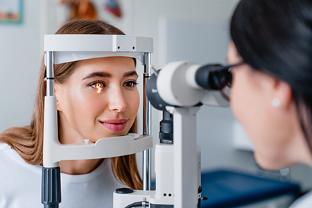
Slit-lamp examination – A magnified, close-up inspection of the front and interior structures of the eye to detect inflammation, infection, or structural changes.
Peripheral visual field testing – Screens for blind spots and peripheral field loss that can indicate glaucoma or neurological conditions.
Intraocular pressure measurement – A quick check for elevated pressure, which is one important risk factor for glaucoma.
Pupil dilation – Dilating drops allow a full view of the retina and optic nerve. This step is especially valuable for detecting retinal disease and vascular changes, and it can add important diagnostic clarity for patients at risk.
Please note: dilating eye drops take about 20 minutes to work, and your eyes may be sensitive to light for a few hours following your exam. It’s wise to bring sunglasses to your visit or have someone drive you home from the exam.
When additional evaluation is necessary, we may recommend diagnostic imaging such as fundus photography, OCT, fluorescein angiography, corneal topography, or automated visual field testing. These modalities capture detailed structural and functional information and are particularly useful for monitoring progressive conditions.
At the practice, our clinicians combine these tools with clinical judgment to create a personalized care plan. Whether the goal is updating a prescription, managing dry eye, monitoring a retinal condition, or coordinating care for a systemic disease with ocular manifestations, we focus on clarity, comfort, and long-term preservation of vision.

Refractive errors are among the most common reasons people seek eye care. They occur when the eye does not focus light precisely on the retina, producing blurred or distorted vision. Refractive errors can develop at any age and are influenced by eye shape, corneal curvature, and changes in the lens.
Symptoms of refractive error include blurry vision at certain distances, headaches, eye strain, or difficulty focusing after prolonged near work. A comprehensive exam identifies the specific type and degree of refractive error so we can recommend the most appropriate correction.
Corrective options range from eyeglasses and contact lenses to refractive surgery when appropriate. Our team discusses the advantages and limitations of each approach and helps patients select a solution that fits their lifestyle, visual needs, and ocular health.
Myopia (nearsightedness) – Objects in the distance appear blurry because light focuses in front of the retina. Myopia often develops in childhood and can progress through the teenage years.
Hyperopia (farsightedness) – Near tasks may be difficult when the eye focuses light behind the retina. Some people compensate by exerting extra focusing effort, which can lead to fatigue or headaches.
Astigmatism – An uneven curvature of the cornea or lens causes distorted or doubled images at any distance. Astigmatism frequently occurs along with myopia or hyperopia.
Presbyopia – With age, the lens becomes less flexible, making near tasks harder; most adults notice this change in their 40s. Reading glasses, multifocal lenses, or specific contact lens designs can restore comfortable near vision.
Detecting a refractive error or an ocular condition is the first step; determining the best path forward is the next. For simple refractive needs, the clinician will refine your prescription and discuss lens options that address visual goals and daily demands. Properly fitted eyewear and contact lenses significantly improve comfort and performance.
When medical or surgical treatment is indicated — for example, managing glaucoma, treating retinal disease, or evaluating candidacy for refractive surgery — the exam provides the diagnostic baseline that guides those interventions. We work collaboratively with ophthalmic surgeons and other specialists when co-management is in the patient’s best interest.
Follow-up intervals are individualized. Some patients benefit from annual exams, while others with higher risk or active disease will be seen more frequently. The goal is to maintain vision stability and intervene early if new concerns arise.

Our visual needs evolve with age, and so do the priorities of each eye exam. For adults approaching middle age, a baseline exam near age 40 helps identify early signs of conditions such as glaucoma, macular degeneration, and changes related to systemic health. Early detection at this stage allows interventions that can slow progression and preserve quality of life.
Older adults face a higher likelihood of cataracts, macular degeneration, diabetic eye disease, and other age-related changes. Even when vision seems stable, routine exams are essential because many of these conditions are treatable or manageable when caught early. We emphasize prevention, monitoring, and practical strategies to maximize independence and visual comfort.
At the other end of the age spectrum, children require age-appropriate testing to ensure proper visual development. Early exams identify issues that can affect learning, hand-eye coordination, and social development. When vision problems are found in childhood, timely treatment often leads to excellent outcomes.

Healthy visual development in children depends on early detection and appropriate care. Professional guidelines recommend exams at several key ages — as an infant, again during the preschool years, before first grade, and then regularly thereafter — though the schedule can vary when risk factors are present.
Comprehensive early exams assess visual acuity, binocular vision (how the eyes work together), tracking, focusing skills, and common pediatric conditions such as amblyopia (lazy eye), strabismus (eye turn), and refractive errors that can interfere with learning.
When treatment is needed, options include eyeglasses, patching or vision therapy, and periodic monitoring. The sooner a problem is identified and addressed, the better the chance for a full functional recovery and normal visual development.
Summary: A comprehensive eye exam is a proactive investment in your vision and overall health. It provides a complete picture of how your eyes work, screens for disease, identifies correctable vision problems, and creates a baseline for future care. To learn more about what to expect or to schedule an exam, please contact us for more information.
A comprehensive eye exam is a detailed evaluation of vision and ocular health that goes beyond a simple vision screening. It assesses visual acuity, how the eyes work together, eye movement and focusing, and the health of internal structures such as the retina and optic nerve. The exam also collects medical history and lifestyle information so clinicians can interpret findings in the context of your overall health.
During the visit, clinicians use a combination of clinical tests and, when indicated, imaging to create a baseline record that can be compared over time. Early detection of subtle changes allows for timely management of eye disease and coordination with other health care providers when systemic conditions are suspected. At Harvard Square Eye Care we use these baseline records to guide personalized care and monitoring plans for each patient.
Frequency depends on age, medical history, and risk factors; there is no single interval that fits everyone. Healthy adults with no symptoms often benefit from exams every one to two years, while people with diabetes, a family history of eye disease, or other risk factors may need more frequent monitoring. Children and older adults typically follow a more proactive schedule because visual development and age-related conditions require closer observation.
Your clinician will recommend a follow-up interval based on test results, risk assessment, and any ongoing treatments or corrective needs. If a new symptom arises between scheduled visits—such as sudden vision changes, flashes, or persistent eye pain—you should contact the practice for prompt evaluation. Regular exams create a continuity of care that improves the ability to detect progressive problems early.
A comprehensive visit includes a series of standard, noninvasive tests designed to evaluate different aspects of vision and eye health. Common components include visual acuity testing, refraction to determine prescription needs, ocular motility and stereopsis testing for eye coordination, pupil response checks, slit-lamp examination of the front structures, and intraocular pressure measurement. These tests provide objective and subjective information that together form a complete picture of visual function.
When indicated, clinicians augment the exam with diagnostic imaging and functional testing such as retinal photography, optical coherence tomography (OCT), corneal topography, and automated visual field testing. Results from these modalities are stored in the chart so changes can be tracked precisely at follow-up visits. Your clinician will explain which tests are performed and why they are relevant to your specific concerns.
Pupil dilation provides a much wider view of the retina and optic nerve than is possible with undilated pupils, making it easier to detect retinal disease, vascular changes, and early signs of conditions such as macular degeneration or diabetic retinopathy. Dilation involves a few drops that temporarily enlarge the pupils, allowing the clinician to examine the internal structures of the eye thoroughly. The procedure is quick, safe, and often essential when the retina or optic nerve needs close inspection.
After dilation you may experience light sensitivity and blurred near vision for a few hours, so it is wise to bring sunglasses and plan for a short recovery time. Driving immediately after dilation can be uncomfortable for some patients, so consider arranging a ride if you expect significant sensitivity. Your clinician will advise whether dilation is necessary for your visit and explain how long effects are likely to last.
The eye offers a unique window into vascular and neurological health because blood vessels and nerve tissue are visible during a retinal exam. Changes in the retina can reflect systemic conditions such as diabetes, hypertension, and certain autoimmune or neurological disorders, sometimes before symptoms appear elsewhere. Identifying these signs enables earlier coordination with primary care or specialty providers for comprehensive management.
During the exam clinicians review your medical history and medications to interpret ocular findings in the proper clinical context, and they may recommend additional testing or referral when systemic disease is suspected. High-resolution imaging and comparison to prior studies improve the accuracy of these assessments. Early detection through routine eye care therefore plays an important role in overall health surveillance.
Bring any current eyewear, including glasses and contact lenses, as well as a list of medications and relevant medical history to help the clinician understand factors that affect your vision. If you wear contact lenses, bring your lens case and follow any instructions provided about temporary discontinuation for certain tests or fittings. Also prepare to discuss your daily visual tasks, screen time, work requirements, and any symptoms you have noticed so the exam can be tailored to your needs.
Plan for an appointment that can last up to an hour, and allow additional time if advanced imaging or dilation is needed. If dilation is likely, bring sunglasses and consider arranging transportation if light sensitivity or blurred near vision would be a problem. Clear communication about your symptoms and history ensures a more focused exam and a practical care plan.
Exams for children are age-appropriate and emphasize visual development, binocular vision, and early detection of conditions that can affect learning and coordination. Testing methods are adapted for developmental level, using objective techniques such as retinoscopy, autorefraction, and binocular vision assessments when subjective feedback is limited. Pediatric exams screen for amblyopia, strabismus, and refractive errors that are most effectively treated when identified early.
For adults the focus often includes monitoring for age-related conditions such as cataracts, glaucoma, and macular degeneration, in addition to addressing refractive needs and occupational visual demands. Baseline evaluations in early middle age help detect subtle changes that may indicate progressive disease. Across all ages, timely intervention and appropriate follow-up are key to preserving long-term visual function.
If you are interested in contact lenses the clinician will perform additional measurements such as keratometry or corneal topography to determine corneal curvature and lens fit. A contact lens evaluation includes a refraction for accurate vision correction, assessment of tear film and ocular surface health, and trial lenses as needed to evaluate comfort and visual performance. Proper fitting reduces the risk of complications and improves day-to-day comfort and vision quality.
Follow-up visits are typically scheduled to confirm fit and to adjust lens parameters, especially for first-time wearers or when changing lens types. The clinician will review safe wear and care practices and recommend an appropriate replacement schedule based on lens material and your eye health. Ongoing monitoring protects the ocular surface and ensures lenses continue to provide optimal vision and comfort.
Advanced imaging technologies, such as optical coherence tomography (OCT), fundus photography, corneal topography, and automated visual field testing, provide high-resolution structural and functional information that complements clinical exam findings. OCT offers cross-sectional views of retinal layers and is invaluable for diagnosing and monitoring macular and optic nerve disorders. Fundus photography documents the retina for comparison over time, while corneal mapping aids contact lens fittings and refractive planning.
These modalities are particularly useful for tracking progressive conditions, assessing treatment response, and establishing precise baselines for long-term follow-up. Images and measurements are saved in the patient record to detect subtle changes that might not be apparent on a single visit. When indicated, your clinician will explain the purpose of each test and how results influence the recommended care plan.
Follow-up intervals are personalized based on the exam findings, risk factors, and any active treatments or monitoring needs. Patients with stable vision and no risk factors may be scheduled at routine intervals, while those with progressive disease, recent interventions, or systemic conditions affecting the eye will have more frequent visits to ensure timely management. The goal is to maintain vision stability and intervene early if new concerns arise.
Imaging and measurement data collected during the initial exam create a baseline that clinicians use to detect subtle changes at subsequent visits and to adjust care as needed. When co-management with ophthalmic surgeons or other specialists is appropriate, the practice coordinates care to ensure continuity and clarity of treatment goals. Harvard Square Eye Care emphasizes long-term monitoring and clear communication so patients understand their follow-up plan and the reasons behind it.

Quick Links
Contact Us